Epidermoid cysts dermoid cysts and teratomatous cysts.
Epidermoid cyst floor of mouth radiology.
Epidermoid cysts are lined by simple squamous epithelium and are more frequently seen in the floor of mouth than in the submandibular space 13.
Napaki and abdul rahman abualruz journal qatar medical.
Dermoid cysts are cysts filled with sebum like material with evidence of specialized skin derivatives 1.
Epidemiology for reasons that are unclear they appear to be more common in the maori of new zealand.
Ranulas are rare benign acquired cystic lesions that occur at the floor of the mouth as sublingual or minor salivary gland retention cysts.
This case reports a 43 year old male patient who presented with a.
Radiology histopathology correlation author salman mirza and shaima fadl and s.
Epidermoid cysts of the floor of the mouth are rare lesions and are much less common than dermoid cysts in the head and neck.
A 13 year old child developed fever and headache.
Epidermoid cysts of the floor of the mouth are rare lesions and are much less common than dermoid cysts in the head and neck.
Epidermal inclusion cysts or epidermal cysts are common cutaneous lesions that represent proliferation of squamous epithelium within a confined space in the dermis or subdermis.
Case report of complicated epidermoid cyst of the floor of the mouth.
Radiology histopathology correlation article mirza2014casero title case report of complicated epidermoid cyst of the floor of the mouth.
Cysts on the floor of the mouth are classified into three types.
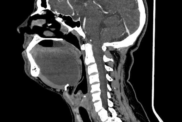
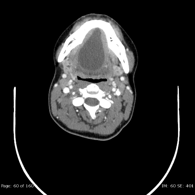
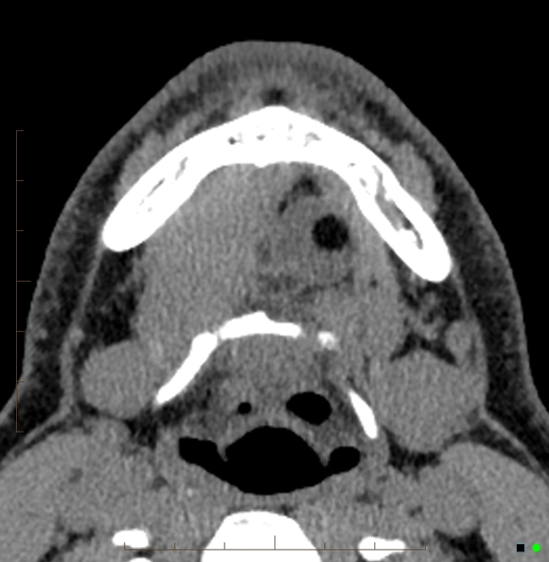
Clinically a cystic midline swelling moving with deglutition was palpable in the submental region with suspicious extension into sublingual space.
This case reports a 43 year old male patient who presented with a longstanding midline swelling in the submental region.
Epidermoid cyst in the floor of the mouth of a 55 year old man.
Dermoid cysts are more common in the head and neck region with the presence of fat content makes it easy for differentiation from an epidermoid cyst.
Dermoid cysts contain skin appendages and cystic teratomas present tissue originating from all the germinal layers.
Floor of mouth dermoid cysts account for 1 6 of all dermoid cysts 2 and they usually present as a midline symmetrical slowly enlarging lesion.
Each type is lined by an epithelial layer but each type exhibits different histologic patterns.
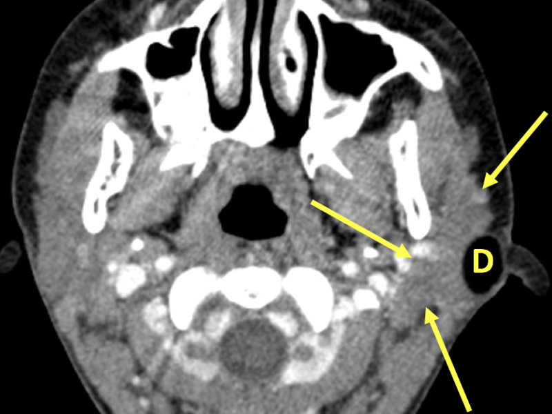
At imaging they appear as midline simple cystic lesions and are often indistinguishable from ranulas fig 12.